
How to Choose Bone Plates in 2026?
How to Choose Bone Plates in 2026? The question is no longer about selecting the strongest implant. It is about matching fixation strategy, anatomy, biology, and clinical evidence. Grand View Research estimates that the global orthopedic implants market reached approximately USD 47 billion in 2023. Fortune Business Insights reports similar growth across trauma and orthopedic devices. These figures show a expanding market, but they do not prove that every new Bone Plates system improves patient outcomes.
Clinical judgment remains essential. Stephan Perren, a leading AO fracture-fixation researcher, offered a clear principle: “The plate is an internal splint.” That sentence still matters. A plate must support healing without unnecessarily damaging blood supply or soft tissue. In 2026, surgeons and procurement teams should examine plate geometry, hole configuration, locking performance, titanium or stainless-steel options, radiographic visibility, and instrument compatibility. They should also review independent clinical studies, fatigue testing, sterilization instructions, and post-market safety data.
Details matter.
A low-profile plate may reduce prominence near the ankle. A stronger construct may be necessary around a weight-bearing joint. However, “stronger” is not automatically better. Excessive stiffness can alter load transfer and influence callus formation. Manufacturer claims require careful verification through published evidence and regulatory documentation. Standards such as ISO 10993 and relevant ASTM testing can support material and mechanical assessments, but they cannot replace patient-specific planning. No checklist is perfect. The most responsible choice combines surgeon experience, transparent data, hospital resources, and the patient’s actual fracture pattern.
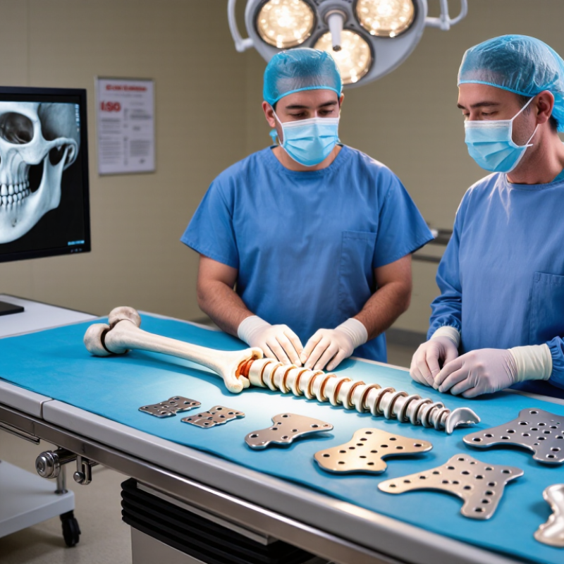
Define the Fracture: AO/OTA Classification and Patient-Specific Risk Data
Choosing a bone plate in 2026 should begin with a precise fracture definition, not a catalogue image. The AO/OTA classification records the bone, segment, fracture morphology, displacement, and comminution. A simple, stable fracture may need a different construct from a multifragmentary or articular injury. Review anteroposterior and lateral radiographs carefully. Use computed tomography when joint involvement or hidden fragments could change the plan. Small details matter.
Patient-specific risk data then guides fixation strength and surgical strategy. Record age, bone density, smoking status, diabetes, kidney disease, medication use, and expected activity level. Osteoporotic bone may require broader load distribution and cautious screw placement. A heavy worker may create higher cyclic forces during recovery. Poor skin condition, swelling, or previous scars can also influence plate position and timing. The safest choice fits both the fracture and the patient.
Do not treat the classification as perfect. Real fractures can sit between categories, and imaging may underestimate instability. Intraoperative findings sometimes change the plan. Experienced surgeons should compare the proposed construct with current clinical evidence, local protocols, and the patient’s healing risks. Mechanical goals should remain clear: restore alignment, protect the joint, preserve soft tissue, and provide enough stability for safe rehabilitation. More metal is not automatically better. 菲律宾申博
How to Choose Bone Plates in 2026? — Define the Fracture: AO/OTA Classification and Patient-Specific Risk Data
A structured framework for matching fracture morphology, mechanical requirements, biological conditions, and patient-specific risk factors before selecting a bone plate construct.
| Assessment Dimension | Classification or Data Point | Clinical Definition | Implication for Plate Selection | Patient-Specific Risk Signal | Recommended Planning Focus |
|---|---|---|---|---|---|
| Fracture location | Bone and segment | Record the affected bone, proximal or distal segment, diaphysis, metaphysis, and articular extension using the AO/OTA system. | Choose a construct that matches the available bone corridor, segment geometry, soft-tissue envelope, and proximity to joints. | Metaphyseal and peri-articular fractures often provide less surface for screw purchase and may have greater fixation complexity. | Review orthogonal radiographs and CT when articular involvement, comminution, or fracture orientation is unclear. |
| AO/OTA fracture group: A | Extra-articular or simple pattern | For many long-bone end segments, Group A describes an extra-articular fracture. In shaft regions, Group A generally represents a simple fracture pattern. | Compression plating may be appropriate for a simple, reconstructable fracture when anatomical reduction and interfragmentary compression are required. | Usually lower geometric complexity than Groups B or C, provided soft-tissue injury and bone quality are favorable. | Assess fracture obliquity, length, contact area, and whether compression would shorten or malalign the limb. |
| AO/OTA fracture group: B | Partial articular or wedge pattern | For end segments, Group B commonly represents a partial-articular fracture. In shaft regions, it generally represents a wedge fracture. | Use a construct that restores the key articular or cortical fragment while preserving blood supply and avoiding excessive compression of intermediate fragments. | Intermediate fragments and partial joint involvement increase reduction and stability demands. | Determine whether the wedge is sufficiently vascularized and whether bridge fixation is preferable to fragment-by-fragment reconstruction. |
| AO/OTA fracture group: C | Complete articular or multifragmentary pattern | For end segments, Group C represents complete articular involvement. In shaft regions, Group C generally represents a multifragmentary fracture. | Usually requires anatomical restoration of the joint surface when involved, combined with a stable metaphyseal or diaphyseal bridge construct. | High mechanical and biological complexity because of comminution, bone loss, soft-tissue trauma, and reduced cortical contact. | Prioritize length, alignment, rotation, joint congruity, soft-tissue preservation, and controlled construct stiffness. |
| Fracture stability objective | Absolute stability | Construct stability intended to eliminate interfragmentary motion at the fracture line, usually through compression and precise reduction. | Consider compression principles for simple fractures where direct healing is desired and the fracture can safely tolerate compression. | Excessive compression can compromise blood supply or shorten a limb if applied inappropriately. | Confirm that the fracture pattern is simple enough for compression and that reduction will not sacrifice alignment or soft-tissue biology. |
| Fracture stability objective | Relative stability | Controlled interfragmentary motion is permitted while length, alignment, and rotation are maintained; healing generally occurs through callus formation. | Bridge plating is commonly considered for comminuted, segmental, or biologically vulnerable fractures where intermediate fragments should not be stripped. | Failure risk rises if the construct is too stiff, too flexible, too short, or exposed to excessive cyclic loading. | Plan plate span, working length, screw distribution, and load sharing according to fracture gap and bone quality. |
| Bone quality | DXA T-score ≤ −2.5 | In adults, a bone mineral density T-score of −2.5 or lower at an accepted measurement site meets the densitometric definition of osteoporosis. | Consider fixation strategies that improve purchase and reduce concentrated stress, such as longer constructs, optimized screw distribution, and carefully selected fixed-angle support when indicated. | Lower bone density may reduce screw holding strength and increase risks of pull-out, loss of reduction, and peri-implant fracture. | Evaluate fracture mechanism, prior fragility fracture, DXA results when available, vitamin D and calcium status, and need for osteoporosis management. |
| Smoking status | Current tobacco use | Smoking is a modifiable biological risk factor associated with impaired bone healing and higher postoperative complication risk. | Do not compensate for poor biology solely by increasing construct rigidity; combine sound fixation with smoking cessation and optimization of healing conditions. | A large orthopaedic meta-analysis reported approximately a 2.5-fold higher risk of nonunion in smokers compared with non-smokers, although the exact risk varies by fracture and procedure. | Document cigarettes or nicotine exposure, provide cessation support, and monitor healing more closely when risk is elevated. |
| Diabetes mellitus | Controlled or uncontrolled diabetes | Diabetes can affect immune function, microvascular perfusion, infection risk, and bone healing; risk depends on disease control and comorbidity burden. | Use biologically respectful approaches, avoid unnecessary soft-tissue disruption, and ensure the construct is appropriate for delayed healing risk. | Poor glycemic control is associated with increased infection and healing complications; no single universal risk percentage applies to every fracture. | Review HbA1c, glucose management, vascular status, renal function, neuropathy, and perioperative infection prevention. |
| Open fracture severity | Gustilo–Anderson I, II, III | Open fractures are graded by wound size, soft-tissue injury, contamination, and vascular or segmental injury. Type III injuries represent the most severe category. | Plate selection must be integrated with debridement, soft-tissue coverage, contamination control, and the overall staged fixation plan. | Higher-grade open injuries have greater infection, nonunion, and soft-tissue failure risks than lower-grade injuries. | Record contamination, vascular status, debridement timing, tissue viability, antibiotic plan, and whether temporary stabilization is required. |
| Soft-tissue envelope | Closed, compromised, or severely damaged | Assess swelling, fracture blisters, skin viability, muscle injury, compartment status, and the planned surgical approach. | Choose an approach and plate position that minimize additional tissue injury; minimally invasive bridge techniques may be considered when appropriate. | Severe soft-tissue damage increases infection risk and may delay definitive fixation or wound closure. | Coordinate fixation timing with swelling, compartment assessment, wound condition, and reconstructive soft-tissue needs. |
| Anatomical reduction requirement | Articular surface involved | Intra-articular fractures require restoration of joint congruity, while extra-articular fractures primarily require correct length, alignment, and rotation. | Articular fractures generally require a strategy that supports the joint surface and maintains reduction under functional loading. | Residual displacement or incongruity may increase the risk of post-traumatic arthritis and functional limitation. | Use CT-based planning when fracture lines, depression, impaction, or separate articular fragments are difficult to define on plain radiographs. |
| Load environment | Low, moderate, or high functional demand | Consider body weight, activity level, expected weight-bearing, muscle forces, and the time required for biological healing. | High-load environments may require greater working length, adequate plate span, balanced screw placement, and protection from premature overload. | Obesity, high activity, falls risk, or inability to comply with weight-bearing restrictions can increase construct loading. | Define postoperative weight-bearing, rehabilitation capacity, fall prevention, and the likely duration of protected loading. |
| Neurovascular status | Intact or compromised | Document distal pulses, capillary refill, motor and sensory function, compartment findings, and any vascular injury. | Fixation strategy and timing may need modification when vascular repair, nerve injury, compartment syndrome, or urgent limb salvage is involved. | Neurovascular compromise is a limb-threatening signal and may supersede routine plate-selection considerations. | Record findings before and after reduction, obtain urgent specialist input when abnormal, and avoid delaying time-critical treatment. |
| Construct verification | Mechanical and biological checklist | Verify reduction, plate position, screw length, fixation density, working length, alignment, rotation, joint penetration avoidance, and soft-tissue respect. | Final selection should balance stability with preservation of fracture biology rather than maximizing the number of screws or construct stiffness. | Risk is increased by malalignment, inadequate fixation in weak bone, excessive stiffness, poor plate span, or unrecognized infection. | Perform intraoperative imaging, document the fixation rationale, and establish a follow-up plan based on fracture and patient risk. |
Related Posts
-
How to Choose the Best Bone Plates for Your Needs?
-

Top 10 Bone Plates in China for Global Buyers Seeking Quality?
-

10 Essential Tips for Optimal Orthopedic Care You Should Know?
-

2026 How to Master Surgical Procedures for Better Outcomes?
-
How to Choose the Right Joint Prosthesis for Your Needs?
-
China Best Orthopedic Trauma Implant Manufacturers?